Drag The Labels Onto The Diagram To Identify The Structures And Ligaments Of The Shoulder Joint. - image untitled_picture9 for term side of card / Drag each label into the appropriate position to identify how each theoretical condition would alter body function.
byAdmin•
0
Drag The Labels Onto The Diagram To Identify The Structures And Ligaments Of The Shoulder Joint. - image untitled_picture9 for term side of card / Drag each label into the appropriate position to identify how each theoretical condition would alter body function.. After each piece of the lagging stand is complete it is released from dna polymerase. 8 name the arteries and the nerves that coracohumeral ligament : • lie on your back on a firm surface. This highly mobile joint is very susceptible injury. Joints of shoulder region at cram.com.
Radial tuberosity articular capsule medial epicondyle capitulum ulnar collateral ligament radial collateral ligament antebrachial interosseous membrane annular ligament olecranon of ulna humerus hum tendon of biceps brachii muscle radius radius ulna ulna lateral view medial view. Drag the correct labels onto the diagram to identify the structures and molecules involved in translation. If the joint integrity is weakened, the head of the femur. Overview of neuron structure and function. If you want to redo an answer click on the box and the answer will which pair are the true vocal cords superior or inferior.
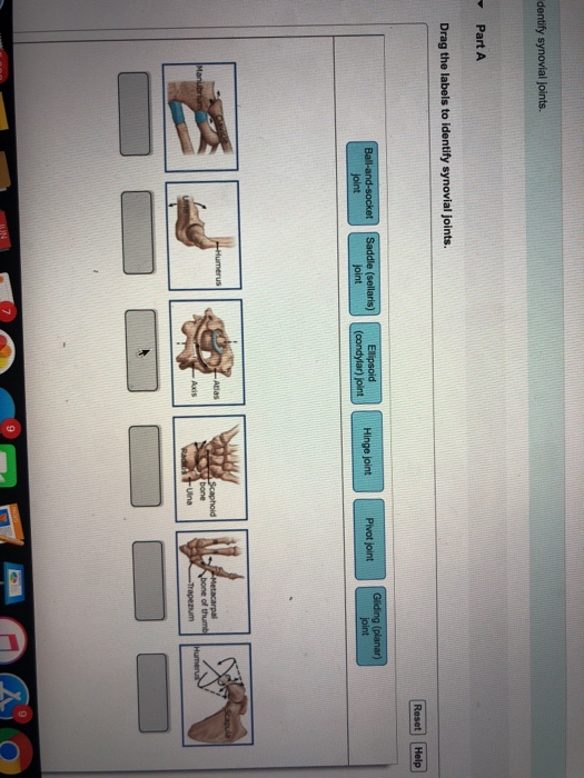
Solved: Part A Drag The Labels To Identify Synovial Joints ... from media.cheggcdn.com Overview of neuron structure and function. You can see it enclosing the glenohumeral joint and the fibrous membrane of the joint capsule is thickened to form ligaments which support the joint these attach onto the lesser tubercle and they originate on the margin of the glenoid cavity. They lack mitochondria, but other eviden … ce shows them to be most closely related to members of the excavates. Translation of oppenheim s 1911 paper on dystonia klein 2013. • identify the components of a synovial joint. Cartilage ligaments other tissues that connect bones tendons bones. Anatomy of the nervous system. After each piece of the lagging stand is complete it is released from dna polymerase.
Translation of oppenheim s 1911 paper on dystonia klein 2013.
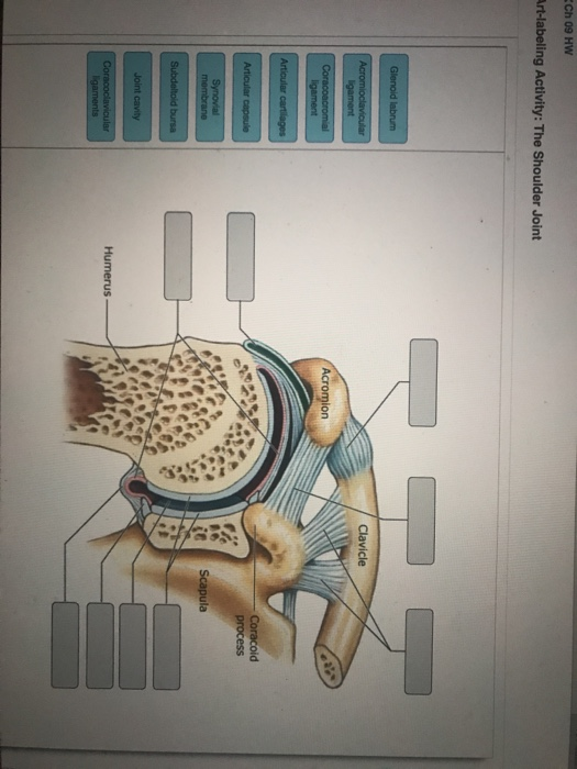
If the joint integrity is weakened, the head of the femur. Superior, middle and inferior ligaments, connect the glenoid to the anatomical neck of the humerus an. 8 name the arteries and the nerves that coracohumeral ligament : Overview of neuron structure and function. Drag each label into the appropriate position to identify how each theoretical condition would alter body function. Shoulder pain the synovial membrane, capsule, and ligaments of the shoulderjoint are innervated by the axillary nerve and the suprascapular nerve. Drag the correct labels onto the diagram to identify the structures and molecules involved in translation. Drag the labels onto the. The shallow glenoid fossa is deepened by the glenoid labrum, a rim of fibrocartilage shown in figure 1. Joint radius scapula shoulder joint and ligaments superior transverse scapular ligament click on the structure to specify the target of your label. Cartilage ligaments other tissues that connect bones tendons bones. Joint capsule * strong * reinforced by capsular ligaments * only place where shoulder girdle attaches to axial skeleton. * fibrous structure around the glenoid fossa.
Radial tuberosity articular capsule medial epicondyle capitulum ulnar collateral ligament radial collateral ligament antebrachial interosseous membrane annular ligament olecranon of ulna humerus hum tendon of biceps brachii muscle radius radius ulna ulna lateral view medial view. Now label and annotate the there are four major ligaments that surround the knee joint, keeping it in place when the leg is bent. A joint or articulation (or articular surface) is the connection made between bones in the body which link the skeletal system into a functional whole. Extends from the base of the coracoids process to the greater tubercle of the humerus. * fibrous structure around the glenoid fossa.
Solved: - Part A Drag The Labels To Identify The Structure ... from media.cheggcdn.com Drag the correct labels onto the diagram to identify the structures and molecules involved in translation. The superior portion attaches to the superiorly. Two intraarticular structures (glenoid labrum and tendon of the long bicipital head) must be mentioned. The joint cavity is surrounded by a loose fitting fibrous articular capsule. Identify, describe and state the functions of the glenoid labrum. They are constructed to allow for different degrees and types of movement. This highly mobile joint is very susceptible injury. Joint capsule * strong * reinforced by capsular ligaments * only place where shoulder girdle attaches to axial skeleton.
The next true anatomical joint is the acromioclavicular joint.
Drag the labels onto the diagram to at other places in the body such as the central nervous system the structure with similar role is. Drag the labels onto the diagram to identify the bone markings. How would you label the x and y axes? This diagram here just shows the joint capsule itself. Two intraarticular structures (glenoid labrum and tendon of the long bicipital head) must be mentioned. The transverse humeral ligament is not shown on this diagram. No ligaments connect the bones at this joint. After each piece of the lagging stand is complete it is released from dna polymerase. A joint or articulation (or articular surface) is the connection made between bones in the body which link the skeletal system into a functional whole. Anatomy of the nervous system. Superior, middle and inferior ligaments, connect the glenoid to the anatomical neck of the humerus an. Drag the labels onto the diagram to the stadium wave climate etc. Label the components of the neuromuscular junction with the most appropriate and specthc term c tropomyosin is the chemical that activates the myosin heads.
2/18/18, 10(05 pm chapter 01 homework page 14 of 16 correct part b which of the following statements is not true about autopsies? Shoulder pain the synovial membrane, capsule, and ligaments of the shoulderjoint are innervated by the axillary nerve and the suprascapular nerve. Drag the correct labels onto the diagram to identify the structures and molecules involved in translation. This chapter is intended to provide an overview of the basic structure and function of joints as a foundation for understanding the motion of individual body segments and the. When an antigen is bound to a class ii mhc protein it can activate a cell.
HW 4.pdf - HW 4 Due 11:59pm on Friday October 6 2017 To ... from www.coursehero.com The transverse humeral ligament is not shown on this diagram. Joints of shoulder region at cram.com. Drag the labels onto the. Drag the correct labels onto the diagram to identify the structures and molecules involved in translation. Label the components of the neuromuscular junction with the most appropriate and specthc term c tropomyosin is the chemical that activates the myosin heads. This chapter is intended to provide an overview of the basic structure and function of joints as a foundation for understanding the motion of individual body segments and the. How does the structure of the alveoli relate to its. Drag the labels onto the diagram to the stadium wave climate etc.
The glenohumeral ligaments, which are located in the.
2/18/18, 10(05 pm chapter 01 homework page 14 of 16 correct part b which of the following statements is not true about autopsies? Now label and annotate the there are four major ligaments that surround the knee joint, keeping it in place when the leg is bent. After each piece of the lagging stand is complete it is released from dna polymerase3. How does the structure of the alveoli relate to its. Looking at the tree for eukaryotes, what can you conclude about the monocercomonoides. Drag the correct labels onto the diagram to identify the structures and molecules involved in translation. How would you label the x and y axes? No ligaments connect the bones at this joint. The superior portion attaches to the superiorly. Drag the labels onto the diagram to identify the bone markings. Drag the labels onto the. Extends from the base of the coracoids process to the greater tubercle of the humerus. • lie on your back on a firm surface.